


TrophyScope office hysteroscopy and TrueBipolar bipolar hysteroresectoscopy
Office hysteroscopy and bipolar hysteroresectoscopy: diagnostics and modern treatmentDo you want to perform a painless examination of the uterus and get an accurate result in one session - without anesthesia, with the minimum diameter of the optics? Schedule a hysteroscopy at Diavita Institute to prevent the development of diseases of the female organs and start treatment in time!
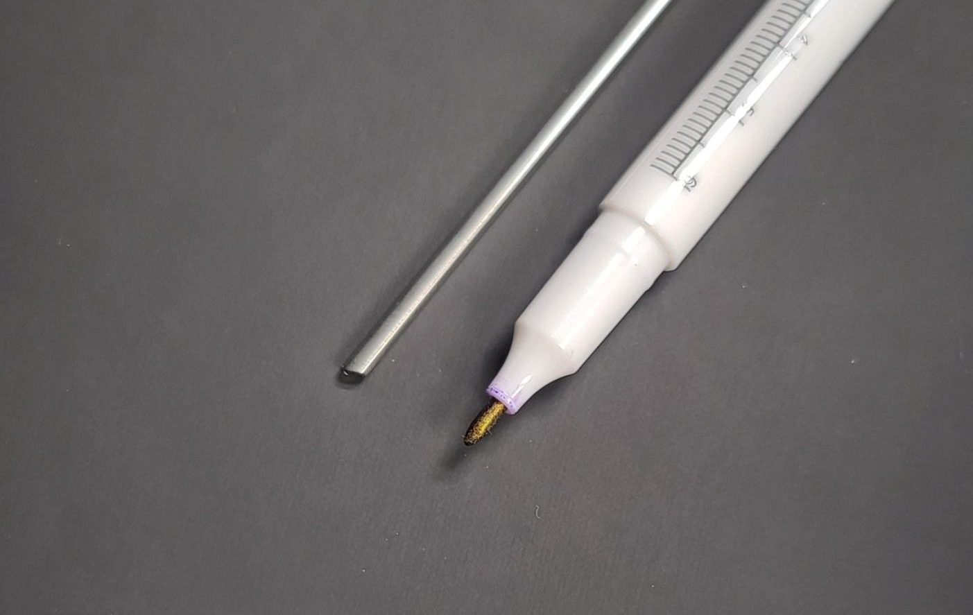
Office hysteroscopy at Diavita Institute is performed using endoscopic optics with a diameter of only 2 mm, that is, our office hysteroscope - TROPHYscope - is three times thinner than usual!
That is why office hysteroscopy is usually a painless outpatient procedure, that is, hysteroscopy without anesthesia. This qualitatively distinguishes office hysteroscopy at the Diavita Institute not only from the outdated scraping technique, but also from ordinary hysteroscopy in Kyiv. The unique endosurgical system from Karl Storz of the TROPHYscope type - a technology developed by the well-known Belgian specialist Rudi L. Campo, MD - not only provides maximum convenience and comfort during office hysteroscopy, but also allows for minimally invasive surgical manipulations - endometrial biopsy, removal of polyps from uterus, dissection of adhesions, coagulation of blood vessels - including the use of extremely safe bipolar energy for resection and coagulation.
Bipolar hysteroresectoscopy using TrueBipolar Plasma technology is an innovative minimally invasive method of diagnosing and treating uterine cavity pathologies. Thanks to bipolar current and plasma technology, the procedure is performed as accurately and safely as possible, with minimal blood loss and a shortened recovery period.
Benefits of TrueBipolar Plasma Hysteroresectoscopy
-
High security: відсутність переходу струму через тіло пацієнтки (як при звичайній монополярній, чи навіть гібридній біполярній резектоскопії), зменшений ризик аритмій.
-
Maximum accuracy thanks to clear high-definition imaging (HD video) and controlled bipolar energy.
-
Minimal thermal trauma surrounding tissues – the plasma current acts only in the treatment area.
-
Reduced blood loss – vascular coagulation occurs instantly during resection.
-
Short rehabilitation period – the patient can return to her usual life.
-
Effective removal even a large amount of pathological tissue in one session.
- Health prevention
Detection and treatment of various problems in the uterine cavity (endometrial polyp, uterine septum, submucosal myoma, etc.). Uterine polyp removal, treatment of submucosal myoma, endometrial atrophic processes, inflammatory changes and others
- Study of the infertility
With the help of office hysteroscopy, it is possible to assess the condition of the uterine cavity, identify possible causes of infertility, such as endometrial polyps, and remove these pathological changes in timely manner
- Treatment of pathology in the uterine cavity
The doctor can take tissue samples (endometrial biopsy) for laboratory analysis, which allows you to get a more accurate picture of the nature of the detected changes, correct pathological changes and restore the healthy state of the uterine cavity
- Psychological comfort
Confidence in your health and the most modern level of diagnosis and treatment
- Detection of pathology
Hysteroscopy makes it possible to detect and eliminate intrauterine pathologies, remove foreign bodies, endometrial polyps, take tissue biopsies, etc. Bipolar resectoscopy is a full range of intrauterine surgery.
- Accurate diagnosis
Hysteroscopy makes it possible to visually visualize the uterine cavity, which allows more accurate diagnosis of various pathologies
- Minimal invasiveness
There is no need for incisions, the minimum diameter of the optics, which reduces the risk of complications
- Security
Office hysteroscopy is performed by experienced doctors according to the method of Professor Rudi L. Campo using the original TROPHYscope equipment and modern technologies from Karl Storz. Hysteroresectoscopy uses the modern technology of bipolar plasma resection - TrueBipolar, which ensures the highest safety and effectiveness.
- Infertility
- Disorders of the menstrual cycle and bleeding
- Abnormalities of the development of the uterus (for example, intrauterine membrane)
- Benign tumors (submucosal myomatous nodes – fibroids)
- Signs of a polyp in the uterine cavity
- Other conditions when it is necessary to visualize and evaluate the uterine cavity
Procedure
During the preliminary consultation, the gynecologist of Diavita Institute determines whether the patient has any complaints, prescribes tests to assess the general state of health. After the consultative and diagnostic appointment, the doctor decides on the conduct of office hysteroscopy or bipolar hysteroresectoscopy in accordance with the available indications. The office hysteroscopy procedure (hysteroscopic polypectomy, diagnostic hysteroscopy, etc.) is performed in full accordance with the latest recommendations and insights from the author of the method - Professor Rudi Campo (Belgium).
Sedation
If necessary, we provide office hysteroscopy with sedation (i.e., controlled administration of sedatives to induce drowsiness or light anesthesia). Sedation can be used to lower pain thresholds, reduce anxiety, and create a comfortable environment for medical procedures. Bipolar hysteroresectoscopy is always performed under general anesthesia.

Duration of the procedure
Office hysteroscopy is an outpatient procedure that is performed at the Diavita Institute by expert doctors as physiologically as possible and usually does not require anesthesia. The duration of the procedure itself is usually less than an hour, after which you can immediately go home or, if necessary, stay for a short time under supervision in the comfortable patient room of the Diavita Institute.

More information about gynecology with comfort and other services

View check-up package offers:

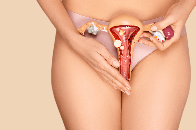
Hysteroscopy is a medical procedure that allows a gynecologist at Diavita Institute to examine the inner surface of the uterus using a special instrument - a hysteroscope. This method is used to diagnose and treat various uterine pathologies, such as polyps, fibroids, anomalies, bleeding, infertility, and others.
The procedure can be diagnostic or therapeutic.
Hysteroscopy is a minimally invasive and effective procedure that allows for accurate diagnosis of many uterine diseases and treatment without large incisions and long recovery times.
Preparing for hysteroscopy:
- Examination before the procedure: the patient is examined by a gynecologist at the Diavita Institute, an ultrasound or tests may be prescribed to assess the condition of the uterus.
- Time of performance: usually hysteroscopy is performed on the 5th-7th day of the menstrual cycle, which allows for an accurate assessment of the condition of its cavity.
- Anesthesia: the procedure can be performed under local anesthesia or under sedation and general anesthesia (especially in the case of hysteroresectoscopy, therapeutic hysteroscopy or in complex cases). Before the intervention, the Diavita Institute doctor may advise taking ibuprofen or diclofenac for a more comfortable procedure. The advantage of office hysteroscopy using the Campo trophoscope is the ability to perform hysteroscopy painlessly and without the use of anesthesia. The advantage of bipolar plasma hysteroresectoscopy TrueBipolar is the highest degree of patient safety when performing the full range of surgical treatment of intrauterine pathology.
The process of conducting:
- Insertion of a hysteroscope: A hysteroscope is a thin tube with a video camera that is inserted through the cervix into the uterine cavity. In the case of a conventional hysteroscopy or hysteroresectoscopy, cervical dilation is used for this, but if the hysteroscopy is performed in an office setting according to Campo, the procedure can be performed without dilation.
- Examination of the uterine cavity: Using a hysteroscope, the doctor can examine the uterus and take photographs or video for further analysis.
- Fluid injection: During the procedure, a sterile solution is injected into the uterus in a controlled manner using a special device, a hysteromat, to perform a complete diagnosis and treatment of intrauterine pathology.
Therapeutic hysteroscopy and hysteroresectoscopy:
If during the diagnosis the doctor detects pathologies (for example, polyps, fibroids, adhesions), it is possible to simultaneously perform therapeutic manipulations, such as:
- Removal of polyps or fibroids,
- Dissection of adhesions,
- Endometriosis treatment,
- Removal of blood clots or other formations.
The optimal and safest treatment method is mechanical (microscissors during office hysteroscopy) and the use of bipolar plasma TrueBipolar electrosurgery.
Duration of the procedure:
Hysteroscopy usually takes between 20 and 60 minutes, depending on the complexity of the case and the required manipulations.
After the procedure:
- After a hysteroscopy, you may experience mild pain in your lower abdomen, similar to menstrual cramps, which may last for several hours or days.
- There may be slight bleeding or vaginal discharge, which is also normal for a few days.
- After the procedure, you should avoid intimate contact and physical exertion for several days or as recommended by your doctor.
Endometrial polyp — is a benign neoplasm on the mucous membrane of the uterus (endometrium), which can be single or multiple. Polyps are often discovered incidentally during a gynecological examination (cervical polyp – cervical canal polyp) or ultrasound. Symptoms of endometrial polyps can vary depending on the size, number, and location of the polyps.
The main symptoms of endometrial polyps:
- Irregular bleeding: intermenstrual bleeding, spotting after intercourse, abnormal menstruation (excessively heavy or prolonged).
- Abdominal pain or discomfort: Mild to moderate pain or discomfort in the lower abdomen, especially before or during menstruation.
- Infertility: Endometrial polyps can cause infertility or difficulty conceiving because they can interfere with the implantation of a fertilized egg or alter normal blood flow to the uterus.
Can uterine polyps be asymptomatic?
Polyps usually do not cause pronounced symptoms and are often only discovered during a medical examination or ultrasound.
When to see a doctor:
- If you notice any of the symptoms listed, especially irregular or abnormal bleeding, it may be a sign of a polyp or other problem, and you should contact your gynecologist at Diavita Institute for advice.
- If you are having difficulty conceiving, or if you are over 40 or have risk factors (e.g., overweight, hypertension, hormonal disorders), be sure to see a doctor for a checkup.
In many cases, endometrial polyps may not cause any symptoms, and the diagnosis is only made during routine gynecological examinations or during an ultrasound examination.
Cervical canal polyp — is usually a benign neoplasm on the mucous membrane of the cervix, which can occur due to chronic inflammation, hormonal disorders, infections or trauma. Polyps may be asymptomatic or cause minor bleeding, pain or discomfort. They are often detected during a gynecological examination or ultrasound at the Diavita Institute.
Methods for removing cervical polyps:
- Curettage (scraping) or mechanical removal: Previously, this was one of the most common methods of removing polyps. A special tool - a curette, forceps or a scalpel - mechanically removed the polyp from the cervical canal. Also, in some cases, it is possible to use electrocoagulation or laser coagulation. All these methods are currently not the method of choice and are not recommended for the treatment of cervical canal polyps because they do not allow the polyp to be removed completely and lead to a high risk of recurrence - because the cervical polyp usually has a long leg, which cannot be removed by these methods.
- Hysteroscopic treatment: It is the method of choice according to modern recommendations. The use of a trophoscope - an office hysteroscope by Campo at the Diavita Institute allows you to fully visualize the cervical canal polyp, determine its full length including the polyp leg, and remove it using safe bipolar surgery. All this is usually possible without anesthesia. Only in some cases is light sedation used. This technique allows for a complete treatment of the cervical canal polyp.
Office hysteroscopy — a quick, minimally invasive method for examining the uterine cavity, diagnosing intrauterine pathology or certain procedures (endometrial biopsy, removal of uterine polyps, etc.).
Bipolar hysteroresectoscopy — is a full-fledged intrauterine operation for the treatment of serious intrauterine pathologies (fibroids, large polyps, developmental anomalies, etc.). The choice of method depends on the diagnosis and clinical situation.
-
The first days: Minor vaginal discharge and mild pain are possible - painkillers and antispasmodics are prescribed to eliminate them.
-
Restoration: Full return to normal activity in 2–3 days; restriction of physical activity and sexual activity for 2–3 weeks.
-
Control inspection: After 7–10 days, assess the condition of the cervix and uterine cavity.
-
Further supervision: Ultrasound in 1–2 months to confirm full recovery.