


Expert ultrasound in Kyiv
Ultrasound is one of the most important diagnostic methods, modern and painlessThere is no radiation exposure when conducting a study at the Diavita Institute, so ultrasound diagnostics is a painless and safe method that does not require the use of contrast agents, is easily accessible, multifunctional, and fast.
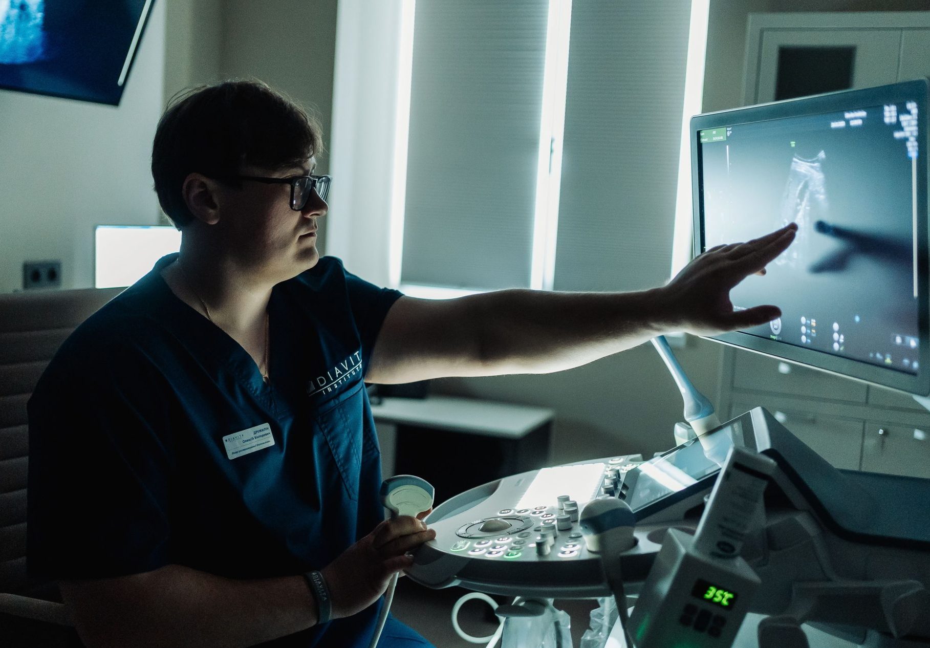
The essence of the ultrasound examination method is that high-frequency sound waves are reflected from internal organs and tissues of different densities, and convert the signals into detailed images on the monitor.
And no, ultrasound is not radiation, so it is completely safe for you!
An ultrasound examination is the best way to confirm or refute the doctor's previous conclusions. The procedure is safe and painless.
Diavita Institute specialists provide an individual approach to each patient. They carefully take all measurements and provide examination results immediately after the examination. When undergoing an ultrasound in Kyiv, you will receive the highest level of service at Diavita Institute.
Ultrasound using 3D and 4D technologies is becoming more and more popular and, at the same time, necessary. Today, this method is very precious diagnostic tool, as it refers to informative and highly accurate methods for examining the pelvic organs and also fetal scan during pregnancy.
Results of 3D/4D ultrasound examination during gynecological ultrasound examination significantly increase the likelihood of making a correct diagnosis, which ultimately affects the patient's management tactics, the appointment of optimal therapy, and therefore the effectiveness of treatment.
During pregnancy, 3D/4D scanning is an additional, but very important method, as its use allows revealing and clarifying some pathological conditions in an unborn child, which reduces the probability of making an incorrect diagnosis.
Color Doppler is a modern technology used by ultrasound machines to display and analyze blood flow in vessels. Allows to simultaneously display the structure of organs and the direction of blood movement, determine its speed and other parameters important for diagnosis.
Types of dopplerometry at Diavita Institute and their advantages:
- Color Doppler:
Allows you to visualize blood flow in the form of colored bands or spots on the image of organs and vessels.
Provides the ability to quickly assess the direction and speed of blood flow without the need for measurement.
- Pulse Doppler:
Allows you to measure blood flow velocity at selected points on the image.
Useful for detecting blood circulation abnormalities and assessing the condition of arteries.
- Spectral Doppler:
Provides detailed information about the speed of blood flow in the form of a spectral diagram.
Allows accurate measurement of various blood flow parameters such as peak velocity and resistance index.
- Power Doppler:
Ideal for visualization of blood flow in small vessels and vascular networks.
It allows detecting even the smallest changes in blood supply, which is important for diagnosing various pathologies.
Doppler is a high resolution, quick results, convenient measurement of blood flow parameters and high diagnostic accuracy. The use of different types of dopplerometry at the Diavita Institute allows doctors to get a complete picture of the blood supply of organs and tissues, which makes this diagnostic method an integral part of modern medical practice.
The Diavita Institute has all the possibilities for providing an expert health assessment using ultrasound diagnostics: first of all, our highly qualified specialists in ultrasound diagnostics, whose level of competence allows performing many unique types of ultrasound examinations at an expert level. And in order for the specialist to reveal and implement his skills, the Diavita Institute has modern professional ultrasound systems, including such as the expert Voluson E10 and Voluson E8 from the leading manufacturer GE Healthcare.
Ultrasound procedures are non-invasive, safe and convenient for our patients. They do not require the use of ionizing radiation and can be carried out without any more complex preparatory measures.
Ultrasound with Comfort
We understand that a visit to the doctor may not be the most pleasant event in the life of every person, so we took care to make it as comfortable as possible for you. No unpleasant sensations. All doctors during the examination communicate with you in detail, show and tell what they see. Any of your questions will not be left without a detailed answer and explanation

Premium equipment
Ultrasound examinations at Diavita Institute are characterized by high accuracy and high detail, as they are performed on top expert equipment - Voluson E8 and Voluson E10, which makes them effective for early diagnosis and study of organ structures.

Dynamic monitoring
Ultrasound diagnostics allows dynamic monitoring of changes in organs and tissues, which is important for the treatment and control of certain conditions
Ultrasound diagnostics is a safe and non-invasive method, without the use of ionizing radiation, which allows obtaining detailed images in real time.
No, ultrasound examinations are completely painless and do not require the use of needles or other invasive methods. During the diagnosis, the patient usually lies on a comfortable couch or chair. The specialist chooses the appropriate sensor (they are convex, linear, endovaginal, etc.) and starts scanning, while the image of the internal organs and tissues under examination is sent to the monitor.
Usually no special preparation is required. However, for some types of ultrasound scans, certain restrictions in food intake, as well as filling or emptying of the bladder before the procedure may be recommended. This guarantees more accurate results.
Preparation for abdominal ultrasound is important to obtain accurate and diagnostically valuable results. Doctors at Diavita Institute recommend adhering to the following principles:
- Diet before the study
1-2 days before the ultrasound, it is advisable to avoid eating foods that can cause gas formation in the intestines, such as: legumes (beans, peas, lentils), potatoes, cabbage (especially white cabbage), fruits containing a lot of sugar (apples, grapes), carbonated drinks.
It is better to eat light food, for example, rice, boiled meat, low-fat fish.
- Desirable restrictions in food and drink
Do not eat or drink for 8 hours before the examination (if the ultrasound is scheduled for the morning).
If the ultrasound is performed in the middle of the day or after lunch, it is recommended to limit food and drink intake 4 hours before the examination.
You can drink still water.
- Stomach and intestinal preparation
An enema or laxative may be recommended if there are problems with intestinal gas, but in most cases this is not necessary.
- Taking medications
If you are taking medications to normalize digestion, such as activated charcoal or drugs to reduce gas formation (simethicone), they can be taken 2-3 hours before the study.
If you have a chronic illness and take medication on an ongoing basis, be sure to consult your doctor.
- Comfortable clothes
During an abdominal ultrasound, you will need to remove your upper body clothing, so wear something light and comfortable.
These recommendations will help optimize results and make research more effective.
Ultrasound diagnostics are widely used to monitor pregnancy and study fetal development without harming the mother or the baby. Ultrasound examinations are considered safe and are often used with the ALARA principle in mind – As Low As Reasonably Achievable.
The frequency of ultrasound examinations depends on specific circumstances and medical indications. The doctor recommends specific ultrasound scans only as necessary.
Abdominal cavity and retroperitoneal space - pathology of the liver, kidneys, pancreas, pain syndromes in the abdomen, oncological diseases, consequences of improper nutrition, abdominal injuries, gallstone disease.
Reproductive system in women – abnormalities of the uterus (congenital and acquired), endometriosis, myoma, ectopic pregnancy, cysts, neoplasms of the uterus and ovaries.
Glands and soft tissues - pathology of the thyroid gland, mammary glands, lymph nodes.
Ultrasound diagnostics can help detect pathological changes, but in some cases confirmation of the diagnosis may require other diagnostic methods, for example, such as biopsy.
Ultrasound (ultrasound examination) of the abdominal organs is a comprehensive examination performed by specialist doctors of the Diavita Institute:
- Liver — assessment of size, structure, and the presence of changes such as inflammation, fatty degeneration, or tumors.
- Gall bladder — checking for stones, polyps, inflammation, or other abnormalities.
- Spleen — determining the size and structure, checking for possible anomalies.
- Pancreas — studying its shape and size, identifying signs of inflammation or tumors.
- Intestine — assessment of the presence of tumors, inflammatory processes or other pathologies.
Abdominal ultrasound may also include examination of abdominal vessels, such as the abdominal aorta, intestines, and others, depending on the clinical situation.
Breast ultrasound at Diavita Institute is usually recommended after menstruation ends, when the hormonal background is more stable and the mammary gland tissues are less sensitive. This is usually 5-10 days of the menstrual cycle.
This period is optimal for ultrasound, as the mammary glands are less responsive to hormonal changes, which makes it easier to detect possible pathologies, such as cysts, tumors or fibroadenomas. In the middle of the menstrual cycle (during ovulation) and before menstruation, the mammary gland tissues may be more swollen, painful and dense, which complicates accurate examination.
However, breast ultrasound can be performed on any day of the cycle if there are indications or relevant symptoms, such as pain, tightness, or discharge from the nipples – regardless of the phase of the menstrual cycle.
Breast ultrasound is a safe and non-invasive method that can detect pathologies including cysts, fibroadenomas, infections, and some forms of cancer.
Preparing for an ultrasound of the kidneys and bladder is important to obtain accurate and high-quality results. Nephrologists Diavita Institute recommends the following:
Full bladder
- For a bladder examination, you must come with a full bladder. This allows the doctor to better assess its size, shape, and the presence of possible abnormalities.
- How to prepare: 1-1.5 hours before the test, drink about a liter and a half of water (you can drink plain water without gas). Do not urinate before the test so that your bladder remains full.
Food and drink
- It is advisable not to eat heavy food (especially fatty and fried) 6-8 hours before the ultrasound to avoid unnecessary gas formation in the intestines.
- If the examination is performed after lunch, you can drink still water and limit your food intake 4 hours before the procedure.
- If you have a tendency to excessive gas or intestinal problems, your doctor may recommend taking activated charcoal or simethicone a few hours before the test.
Other points
- If you take any medications regularly, especially for chronic conditions, please inform your doctor before the procedure. Your doctor may recommend changing the time of your medication or prescribing other medications during the preparation period.
- Wear comfortable clothing, as you will likely need to undress your upper body for the examination.
These recommendations will help make the study more accurate and comfortable for you.